
All the CT images were taken and reconstructed with a same protocol.

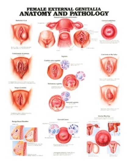
All animals underwent CT examination under general anesthesia and positioned in sternal recumbency. For this purpose, 10 male and 10 female clinically normal, adult intact mixed-breed dogs weighing 15.00 to 20.00 kg were selected randomly. This study aimed to evaluate the normal pelvic diaphragm muscles (levator ani and coccygeus muscles) using the computed tomography (CT) scan. Despite the wealth of information available, many gaps in knowledge remain and will require a community-wide effort to come to a more robust model of female genital evolutionary patterns.Įvaluation of pelvic diaphragm muscles in dogs merits clinical attention because of the anatomical importance and their involvement in perineal hernia. The evidence for either bilobed or unitary glandes clitorides is ambivalent. Based on available information, the ancestral eutherian configuration of the external female genitalia included a cervix, a single vaginal segment, a tubular UGS, and an unperforated clitoris close to the entrance of the genital canal. Some morphs are unique to early branching clades, like the absence of the cervix, while others arose multiple times independently, like the flattening out or loss of the UGS, or the extreme elongation of the clitoris. A preliminary phylogenetic analysis shows two patterns. It includes the absence of whole anatomical units, like the cervix in many Xenarthra, or the absence of the urogenital sinus (UGS), as well as the complete spatial separation of the external clitoral parts from the genital canal (either vagina or UGS). Variation in female external genitalia is anatomically more radical than that in the male genitalia. A review of the literature on the anatomy of the lower female genital tract in therian mammals reveals, contrary to the general perception, a large amount of inter-specific variation.


 0 kommentar(er)
